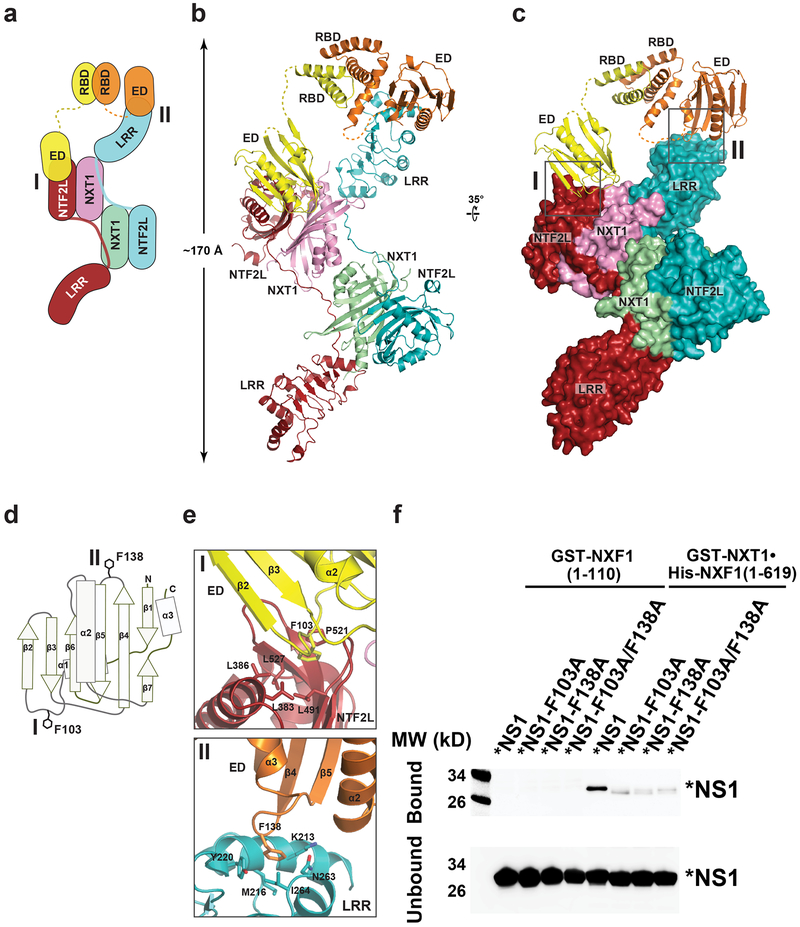
Fig. 2 |. Structure of a 2:2:2 complex of *NS1•NXF1117–619•NXT1.
a, Schematic diagram of the *NS1•NXF1117–619•NXT1 complex. *NS1 binds to NXF1117–619•NXT1 through two interfaces, I and II. b, c, Overall structure of *NS1•NXF1117–619•NXT1 in two orientations, colored as in (a). Insets illustrate the *NS1-NXF1117–619•NXT1 binding interfaces, and are expanded in (e). d, Schematic diagram of the NS1-ED domain. e, Expanded views of Interface I and II, viewed as in (c). f, In vitro GST-Pull down assays with NXF1•NXT1 and * NS1 or *NS1 with mutations on the NXF1 binding site shown in (e). Purified GST-NXT1•His-NXF1 was incubated with purified *NS1, *NS1-F103A, *NS1-F138A, or *NS1-F103A/F138A. NS1 was detected by western blot and showed diminished interaction upon mutations on the NXF1 binding site. The N-terminal domain of GST-NXF1(1–110) was used as control. n=3.

