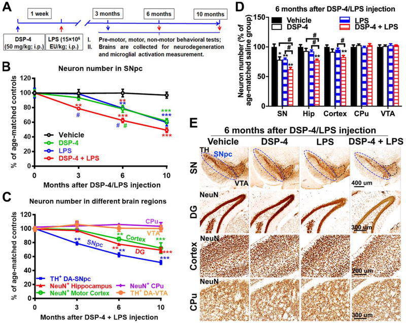
Fig. 1. DSP-4 treatment accelerates neurodegeneration in LPS-treated mice brain.
(A) Schematic drawing of the treatment regimen for DSP-4 and LPS. Male C57BL/6J mice received a single injection of DSP-4 (50 mg/kg, i.p.), followed by LPS (15×106 EU/kg, i.p.) treatment one week later. 3, 6 and 10 months after LPS injection, mice were sacrificed for immunohistological staining of different brain regions. (B) DSP-4 accelerates SN-DA neurodegeneration in LPS-treated mouse brains [two-way ANOVA, treatment x time interaction, F(9,50)=5.525, p<0.0001; effect of time, F(3,50)=49.93, p<0.0001; effect of treatment, F(3,50)=28.36, p<0.0001], *p<0.05, **p<0.01, ***p<0.001 compare with age-matched vehicle controls. #p<0.05 compare with age-matched DSP-4- (green) or LPS- (blue) treated animals. (C) Quantitative analysis of neuron loss in the different brain regions at different time points after “two-hit” treatment. TH-immunoreactive neurons in both SNpc and VTA were counted stereologically. Neurons in hippocampal granule layer (Neu-N-immunoreactive), motor cortex and CPu (Neu-N-immunoreactive) were quantified by auto-counting using ImageJ software. Results are expressed as a percentage of age-matched vehicle controls (mean ± SEM) in each group at each time point [one-way ANOVA for each region with time as main effect, SNpc: F(3,11)=55.07, p<0.0001; Hippocampus: F(3,9)=31.93, p<0.0001; Motor cortex: F(3,19)=8.938, p=0.0007; CPu: F(3,17)=0.2363, p=0.8698; VTA: F(3, 4)=0.3179, p=0.8123], **p<0.01, ***p<0.001 compare with age-matched vehicle controls. (D) Quantitative analysis of neuron loss in different brain regions at 6 months after DSP-4, LPS, or “two-hit” treatment. Hip=Hippocampus. Results are expressed as a percentage of age-matched vehicle controls (mean ± SEM) from 4-5 mice in each group at each time point [two-way ANOVA, treatment x brain region interaction, F(9,89)=3.364, p=0.0014; effect of treatment, F(3,89)=9.949, p<0.0001; effect of brain region, F(3,89)=20.4, p<0.0001]. *p<0.05 and **p<0.01 compare with age-matched vehicle controls. #p<0.05 compare with indicated groups. (E) Representative images of staining in SN, hippocampus, motor cortex, and CPu at 6 months after injection were shown. Dopaminergic neurons in the SNpc were stained with anti-TH antibody. Neurons in hippocampal granule layer, motor cortex and CPu were stained with anti-Neu-N antibody. Scales as indicated in the pictures.

