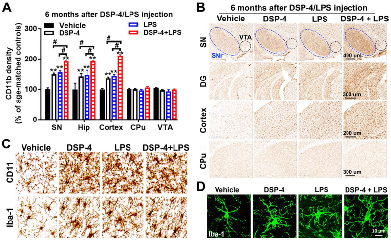
Fig. 2. DSP-4 treatment potentiates microglial activation in LPS-treated mice.
Male C57BL/6J mice received a single injection of DSP-4 (50 mg/kg, i.p.), followed by LPS (15×106 EU/kg, i.p.) treatment one week later. 3, 6 and 10 months following the LPS injection, microglia in SN, VTA, hippocampus, motor cortex and CPu were stained with CD11b antibodies. (A) Quantitative analysis of microglial activation in these brain regions at 6 months after injection by measuring alterations of CD11b density [two-way ANOVA, treatment x brain region interaction, F(12,109)=12.4, p=0.0014; effect of treatment, F(4,109)=69.91, p<0.0001; effect of brain region, F(4,109)=66.77, p<0.0001]. **p<0.01 compare with age-matched vehicle controls. #p<0.05 compare with indicated groups. (B) Representative images of CD11b staining in different brain regions at 6 months after injection were shown. Scales as indicated in the pictures. (C-D) High-power images show the morphological changes in microglia and an increase in microglial CD11b and Iba-1 in SN at 6 months after DSP-4/LPS injection.

