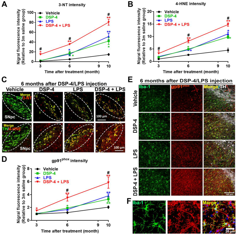
Fig. 3. DSP-4 treatment potentiates neuronal oxidative stress in LPS-treated mice.
Male C57BL/6J mice received a single injection of DSP-4 (50 mg/kg, i.p.), followed by LPS (15×106 EU/kg, i.p.) treatment one week later. 3, 6 and 10 months following the LPS injection, oxidative damage in protein and in lipid in SN were stained with anti-3-NT or anti-4-HNE antibody. (A-B) The fluorescence intensity of 3-NT [two-way ANOVA, treatment x time interaction, F(6,101)=11.28, p<0.0001; effect of treatment, F(3,101)=54.13, p<0.0001; effect of time, F(2,101)= 178.1, p<0.000l] (A) and 4-HNE [two-way ANOVA, treatment x time interaction, F(6,88)=11.1, p<0.0001; effect of treatment, F(3,88)=65.31, p<0.0001; effect of time, F(2,88)=212.2, p<0.0001] (B) in the SN was quantified. (C) Representative images show double labeled TH and 3-NT (top) and TH and 4-HNE (bottom) in SN 6 months after LPS injection. Images were captured at 40× magnification. The SNpc zone is outlined in white dots. (D) The fluorescence intensity of gp91phox in the SN was quantified [two-way ANOVA, treatment x time interaction, F(6,90)=2.382, p=0.035; effect of treatment, F(3,90)=10.11, p<0.0001; effect of time, F(2,90)=42.03, p<0.0001]. (E) SN were triple labeled for gp91phox, Iba-1 and TH. Images were captured at 40× magnification. (F) The high-power images to show co-localization of Iba-1 and gp91phox in SN. The selected SN area was confirmed by TH (white) staining. Results are expressed as a percentage of age-matched vehicle controls (mean ± SEM). *p<0.05, **p<0.01 compare with age-matched vehicle controls. #p<0.05 compare with age-matched DSP-4 and LPS alone groups. Scales as indicated in the pictures.

