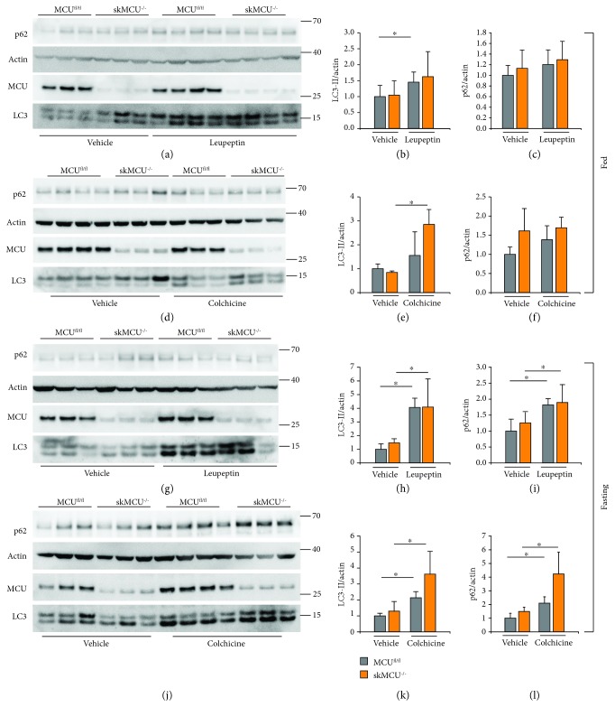
Figure 2.
Autophagy flux analyses in skMCU−/− mice. (a-f) Immunoblot of EDL muscles of skMCU−/− or skMCUfl/fl mice treated or not with leupeptin (a-c) or colchicine (d-f) in fed conditions. Western blot analyses demonstrated efficient MCU deletion in EDL muscles. LC3-II and p62 protein levels were used to monitor autophagy, relative to actin protein levels used as loading control. (b, c, e, and f) Quantification by densitometry of the ratio between LC3-II/actin and p62/actin. ∗p < 0.05, t test (two-tailed, unpaired) of three vehicle animals and four treated animals, respectively. Data are presented as mean ± SD. (g-l) Immunoblotting analysis of EDL muscles of skMCU−/− or skMCUfl/fl mice treated or not with leupeptin (g-i) or colchicine (j-l) upon starvation. Western blot analyses demonstrated efficient MCU deletion in EDL muscles. (h, i, k, and l) Quantification by densitometry of the ratio between LC3-II/actin and p62/actin. ∗p < 0.05, t test (two-tailed, unpaired) of, respectively, three vehicle animals and four treated animals. Data are presented as mean ± SD.

