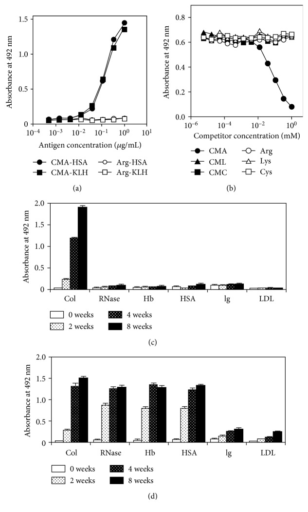
Figure 2.
Immunoreactivity of monoclonal anti-CMA antibody (3F5). (a) Each well coated with the indicated concentration of the test sample was reacted with 3F5 (1 μg/mL). The antibody bound to the wells was detected by HRP-conjugated anti-mouse IgG. (b) Specificity of the anti-CMA antibody (3F5). Each well was coated with 0.1 mL of 1 μg/mL CMA-conjugated KLH and blocked with 0.5% gelatin. 60 μL of 3F5 (2 μg/mL) and 60 μL of the samples were preincubated for 1 h, and sample aliquots (100 μL) were added to each well and incubated for 1 h. The antibody bound to the wells was detected as described above. (c) CMA and (d) CML formations in glycated protein samples. Type I collagen (Col), HSA, RNase, hemoglobin (Hb), immunoglobulin (Ig), and LDL were incubated with 200 mM glucose at 37°C for up to 8 weeks, and the CMA or CML content of the samples was determined by ELISA.

