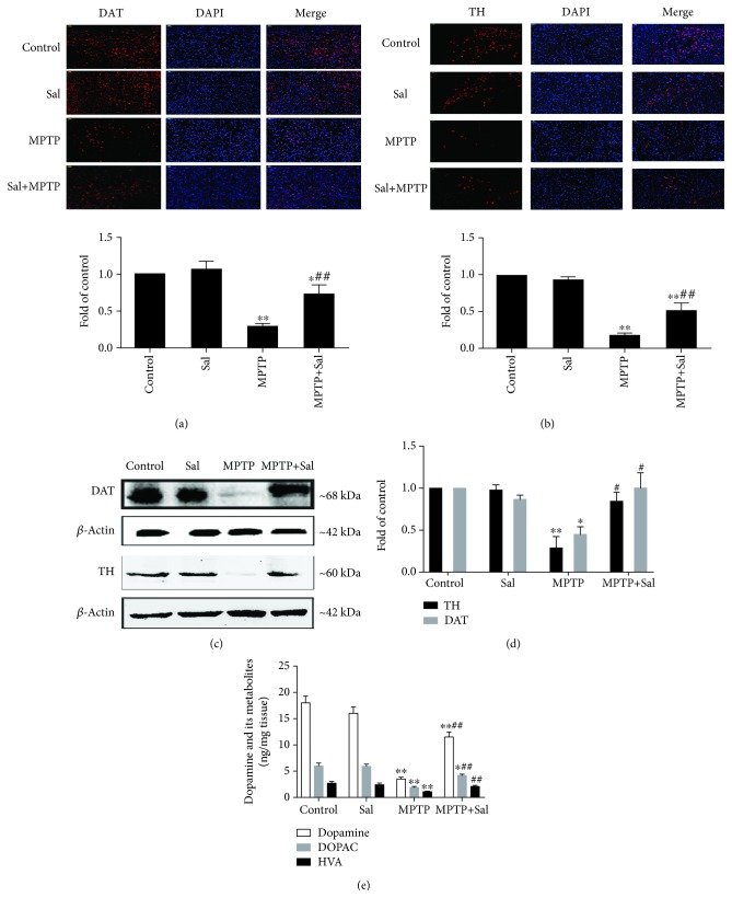
Figure 2.
Effect of Sal on MPTP-induced DA neuron damage. Representative photomicrographs and quantitative analysis of DAT-positive neurons in the striatum (a) and TH-positive neurons in the SN (b). Scale bar, 50 μm. (c) Western blot analysis of DAT expression in the striatum and TH expression in the SN of mice. β-Actin served as loading controls. (d) Qualification analysis of protein expression of DAT and TH in mice. (e) Effect of Sal on the level of dopamine, DOPAC, and HVA in the striatum of mice. Each column represents the mean ± SD (n = 3). ∗P < 0.05 and ∗∗P < 0.01, compared with the control group; #P < 0.05 and ##P < 0.01, compared with the MPTP-treated group.

