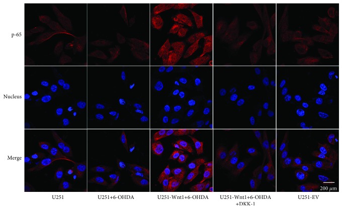
Figure 9.
Immunofluorescence staining of p65 protein in U251 cells. U251 cells, U251-Wnt1 cells, or U251-EV cells were treated with vehicle, 6-OHDA (50 μM), or/and DKK-1 (100 ng/ml) for 24 h, and the expression and distribution of p65 were examined using an antibody against p65 (red). Nuclei were counterstained with DAPI (blue). All images were captured using a confocal laser scanning microscope.

