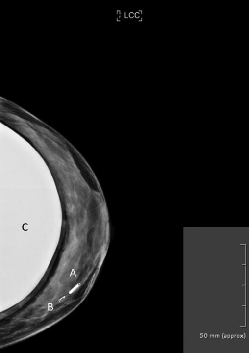
Figure 2. Left breast mammography of a 30-year-old woman. (A) Shows the LOCalizer™ chip which was placed in a non-palpable lesion. Note the direct comparisons in size when compared to a standard titanium clip (B). The patient received a bilateral sup-pectoral augmentation “C” 5 years earlier. In this case the non-palpable breast lesion had been clipped before, however the clip was dislocated. A stereotactic wire marking was also not possible due to the risk of breast implant damage. Thus, a LOCalizer™ chip was placed in the accurate position.

