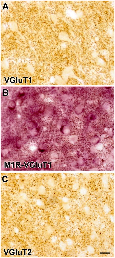
Figure 1.
(A) Photomicrograph of VGluT1 immunoreactivity in the BLa (DAB immunoperoxidase). Note that neuronal somata and proximal dendrites are unlabeled but there are punctate VGluT1+ axons and axon terminals in the neuropil. (B) Photomicrograph of a section stained for M1R with V-VIP as a chromogen in the BLa. Note that somata of putative PNs are stained purple. This section was also stained for VGluT1 immunoreactivity with DAB as a chromogen. The neuropil contains small punctate structures that represent VGluT1+ axons and M1R+ processes. (C) Photomicrograph of VGluT2 immunoreactivity in the BLa (DAB immunoperoxidase). Note that somata and proximal dendrites are unlabeled but there are punctate VGluT2+ axons and axon terminals in the neuropil. Scale bar = 20 μm for A, B, and C.

