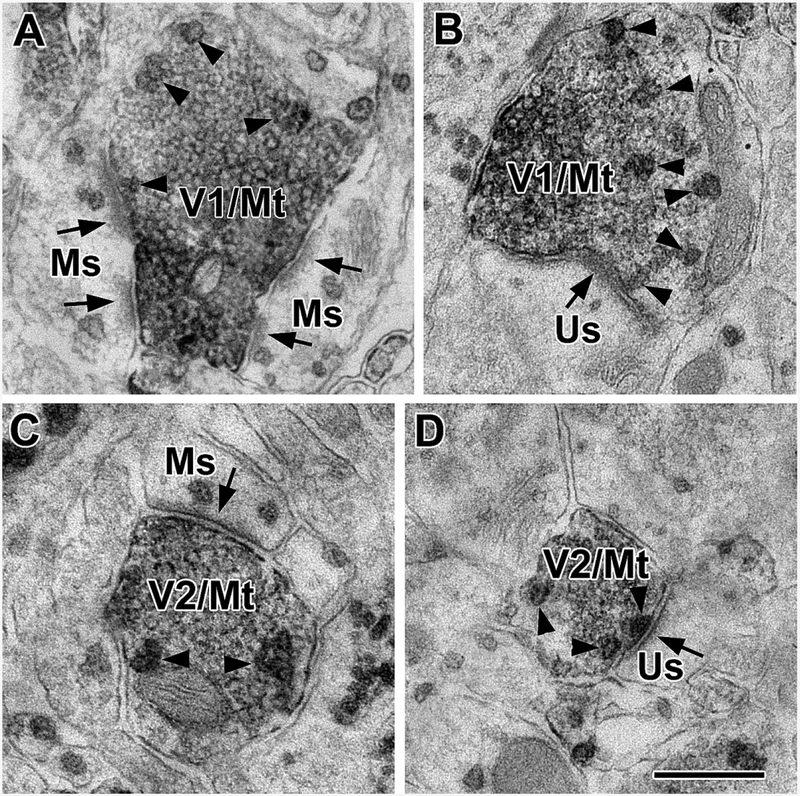
Figure 4.
Terminals immunoreactive for both VGluT1 and M1R (V1/Mt) and terminals immunoreactive for both VGluT2 and M1R (V2/Mt) form asymmetrical synapses with M1R+ spines (Ms) and unlabeled M1R-negative spines (Us) in the BLa. Arrowheads indicate M1R+ particles in terminals. (A) A VGluT1/M1R double-labeled terminal forms asymmetrical synapses with two M1R+ spines. (B) A VGluT1/M1R double-labeled terminal forms an asymmetrical synapses with an unlabeled spine. (C) A VGluT2/M1R double-labeled terminal forms an asymmetrical synapse with an M1R+ spine. (D) A VGluT2/M1R double-labeled terminal forms an asymmetrical synapse with an unlabeled spine. Scale bar = 500 nm.

