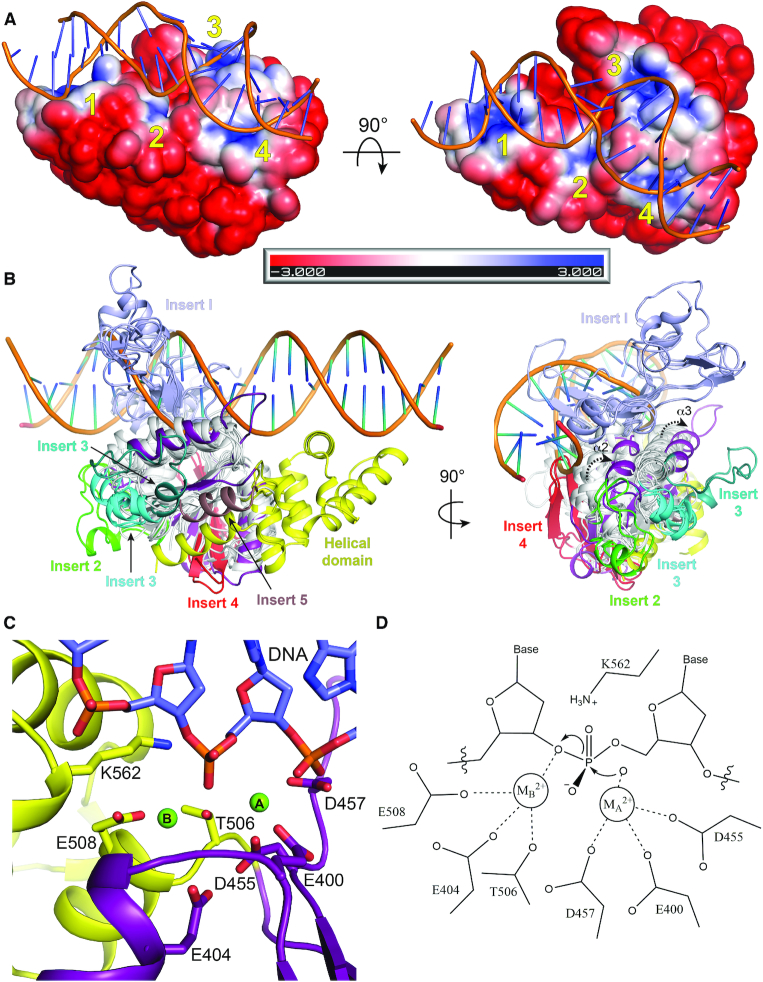
Figure 7.
Helical domain orients DNA binding in Class 2 OLD nucleases. (A) Electrostatic surface of BpCTR with modeled DNA (G:T mismatched substrate taken from PDB: 3K0S). Electrostatic potential calculated with APBS (37). Scale indicates coloring of the potential from –3KbT/ec to +3KbT/ec. The four basic patches around the active site cleft are numbered in yellow. (B) Side and end views of Toprim family superposition with modeled DNA substrate bound to Bp OLDCTR. Toprim cores are colored white with Inserts 1–5 individually labeled. Bp Toprim and helical domains are colored purple and yellow respectively. (C) Arrangement of BpCTR active site with modeled DNA. (D) Proposed two-metal catalysis mechanism for Class 2 OLD nuclease cleavage.

