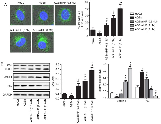
Figure 4.
HF promotes the autophagy of H9C2 cells under AGEs-induced ER stress. H9C2 cells were randomly divided into five groups: Control group, normal H9C2 cells; AGEs group, AGEs-induced cells; and three experimental groups, AGEs-induced cells treated with HF at different concentrations (0.5, 2 and 8 nM) for 24 h. (A) Expression levels of LC3 were detected via immunofluorescence (magnification, ×400). (B) Expression levels of autophagy-associated proteins (LC3, Beclin 1 and P62) were measured by western blotting. Experiments were repeated at least three times, and data are presented as the means ± standard deviation. *P<0.05 vs. the H9C2 group; #P<0.05 vs. the AGEs group. AGEs, advanced glycation end products; GFP, green fluorescent protein; HF, halofuginone; LC3, microtubule-associated proteins 1A/1B light chain 3B.

