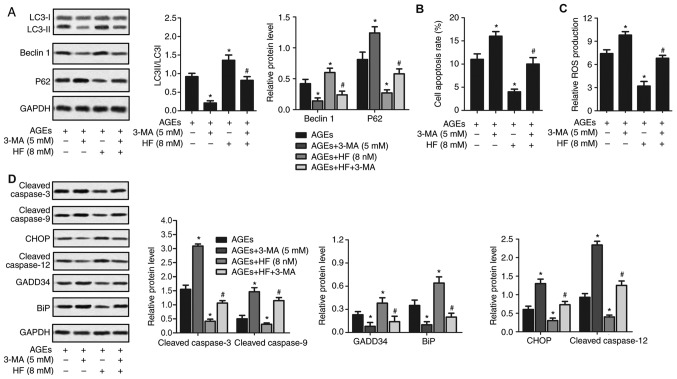
Figure 5.
HF protects H9C2 cells from AGEs-induced damage by inducing autophagy. H9C2 cells were randomly divided into four groups: AGEs group, AGEs-induced cells; AGEs + 3-MA group, AGEs-induced cells treated with 3-MA (5 mM) for 24 h; AGEs + HF group, AGEs-induced cells treated with HF (8 nM) for 24 h and AGEs + 3-MA + HF group, AGEs-induced cells treated with 3-MA (5 mM) and HF (8 nM) for 24 h. (A) Western blot analysis of the autophagy-associated proteins (LC3, Beclin 1 and P62). GAPDH was used as an endogenous reference. Flow cytometry was used to determine (B) cell apoptosis rate and (C) relative ROS production. (D) Expression levels of apoptosis-associated proteins (cleaved caspase-3 and cleaved caspase-9), ER stress-associated proapoptotic proteins (CHOP and cleaved caspase-12) and prosurvival proteins (GADD34 and BiP) were detected via western blot analysis. GAPDH was used as an endogenous reference. Experiments were repeated at least three times, and data are presented as the means ± standard deviation. *P<0.05 vs. the AGEs group; #P<0.05 vs. the AGEs + HF group. 3-MA, 3-methyladenine; AGEs, advanced glycation end products; BiP, binding immunoglobulin protein; CHOP, CCAAT/enhancer-binding protein homologous protein; ER, endoplasmic reticulum; GADD34, growth arrest and DNA damage-inducible protein GADD34; HF, halofuginone; LC3, microtubule-associated proteins 1A/1B light chain 3B; ROS, reactive oxygen species.

