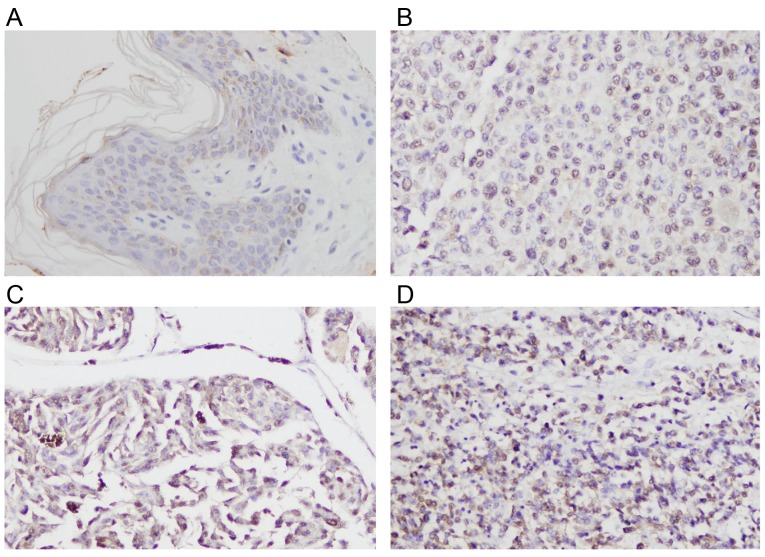
Figure 1.
Expression of Rsf-1 in melanoma tissue. (A) Negative Rsf-1 staining observed in normal skin tissue. Moderate Rsf-1 staining was detected in cases of melanoma tissue of stages (B) II and (C) III. (D) Strong Rsf-1 staining was detected in a case of melanoma tissue in stage IV. Magnification, ×400. Rsf-1, remodeling and spacing factor 1.

