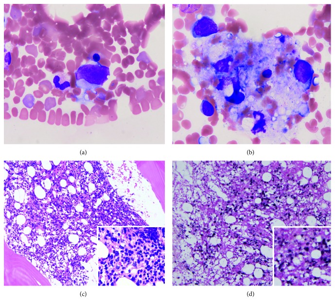
Figure 1.
Bone marrow aspiration/biopsy findings. (a, b) Bone marrow aspirate. Wright-Giemsa staining. (a) A large, atypical lymphocyte (×1,000). (b) Hemophagocytosis (×1,000). (c, d) Bone marrow biopsy showing hypercellular marrow. (c) Hematoxylin and eosin staining. Low-magnification image (×40). Inset: high-magnification image (×200). (d) Epstein–Barr virus-encoded RNA (EBER) in situ hybridization. Large atypical lymphocytes are positive. Low-magnification image (×40). Inset: high-magnification image (×200).

