Figure 2.

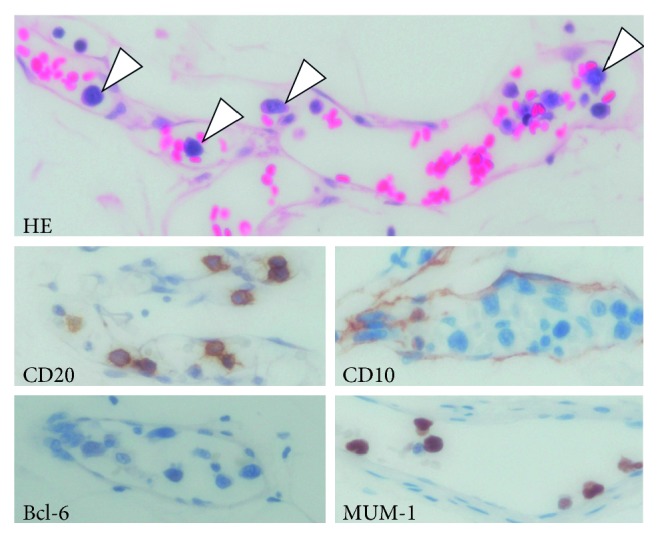
Histopathology of the angiolipoma. Large atypical lymphocytes in the lumina of the capillaries. Lymphoma cells are indicated by arrowheads. They are CD20(+), CD10(−), Bcl-6(−), and MUM-1(+). Magnification, ×400. HE, hematoxylin and eosin stain.
