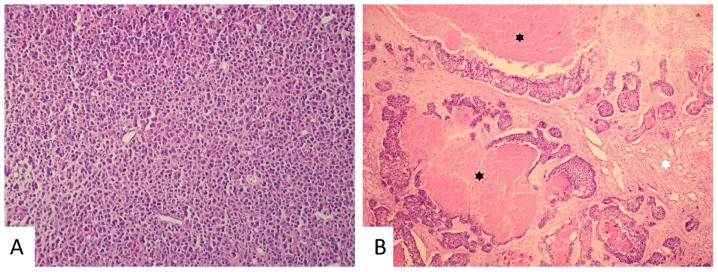
Figure 1.
Representative images of invasive breast carcinoma NST prior and following neoadjuvant chemotherapy. (A) Invasive breast carcinoma NST of a solid architecture in a core needle biopsy prior to therapy. Haematoxylin and eosin staining, magnification, ×200. (B) Residual carcinoma of a predominantly solid architecture with scattered cribriform pattern with regressive changes [focal necrosis (black asterisks) and fibrosis (white asterisk)] following therapy. Haematoxylin and eosin staining, magnification, ×100. NST, no special type.

