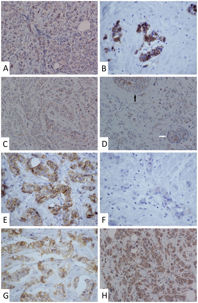
Figure 2.
Representative images of changes in the expression of claudins in invasive breast carcinoma no special type following neoadjuvant chemotherapy. (A) Punctate membranous positivity of claudin-1 in a core needle biopsy prior to therapy. Magnification, ×200. (B) Strong and continuous membrane staining of claudin-1 in the residual cancer from the same patient. Magnification, ×600. (C) Punctate-to-continuous membranous positivity of claudin-1 in a core needle biopsy prior to therapy. Magnification, ×200. (D) Similar staining pattern of claudin-1 in the residual cancer from the same patient. Continuous and intense positivity was observed in ductal carcinoma in situ (black arrow) and in the small residual non-tumour duct (white arrow). Magnification, ×200. (E) Strong and continuous membranous staining of claudin-3 in a core needle biopsy prior to therapy. Magnification, ×600. (F) Faint claudin-3 staining in the residual cancer from the same patient. Magnification, ×600. (G) Moderate membranous positivity of claudin-4 in a majority of the tumour cells in a core needle biopsy before therapy (retraction artefacts). Magnification, ×600. (H) Similar staining pattern of claudin-4 (comparing membranous positivity only) in the residual cancer from the same patient. Magnification, ×200.

