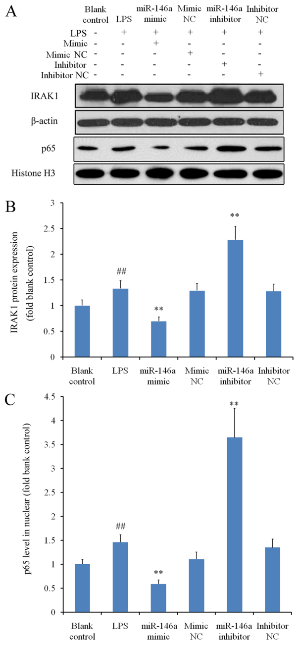
Figure 3.
Inhibitory effects of miR-146a on IRAK1 expression and NF-κB activation under inflammatory conditions. THP-1 cells were treated with LPS following transfection with either a miR-146a mimic or inhibitor to activate the inflammatory signaling response. Western blot analysis was used to measure relative protein expression levels of IRAK1 and nuclear NF-κB p65 subunit, normalized to their respective internal references. Three independent experiments were performed. (A) Representative images of the western blot analysis. Relative protein expression levels of (B) IRAK1 and (C) the nuclear p65 subunit. ##P<0.01 vs. blank control and **P<0.01 vs. corresponding NC groups. miR, microRNA; IRAK1, interleukin-1 receptor-associated kinase 1; NF-κB, nuclear factor κB; LPS, lipopolysaccharide; NC, negative control.

