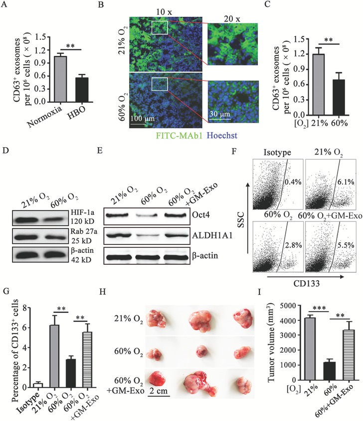
Figure 7.

Respiratory hyperoxia attenuates colon cancer cell stemness through inhibiting GM‐Exo production. A) Exosomes were quantified from tumoral G‐MDSCs treated with HBO or maintained at normoxia in vitro by ExoELISA. B) Immunohistochemical demonstration of 60% O2 treatment reduce hypoxic areas (green) in tumor tissue. Tissue cryosections from intradermal CT‐26 tumors were prepared, and immunohistochemistry was performed. Immunofluorescence images were representative of six random fields. C) Exosomes were quantified from tumoral G‐MDSCs from CT‐26 bearing mice treated with 60% O2 or maintained at 21% O2 by ExoELISA. D) Western blotting was performed to show HIF‐1α and Rab27a protein levels in tumoral G‐MDSCs from CT‐26 bearing mice treated with 60% O2 or maintained at 21% O2. Representative results from three independent experiments. E) Western blotting was performed to show Oct4 and ALDH1A1 protein levels in tumor tissue from CT‐26 bearing mice treated with 60% O2 or maintained at 21% O2. Representative results from three independent experiments. F,G) The percentage of CD133‐positive tumor tissue cells were analyzed by FCM. The data are represented as the mean ± SEM of each group pooled from three independent experiments. H,I) The effect of respiratory hyperoxia on the development of an CT‐26 cell tumor model. Mice with 15‐day established CT‐26 colon tumors were treated with 60% O2, and some of them were treated with 30 µg GM‐Exo via the tail vein once every 3 days from 15‐day. Tumors were harvested at day 25. Representative tumors in each group are shown and the volume of tumors in each group is measured. In A,C) the data are presented as the mean ± SEM of each group pooled from three independent experiments. **p < 0.05, analyzed by a t‐test. In G,I) the data are presented as the mean ± SEM. **p < 0.01,***p < 0.001, analyzed by ANOVA.
