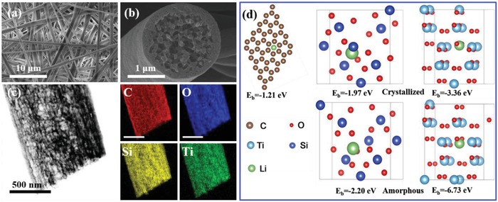
Figure 1.

a) Top‐view SEM images of the top surface of the PCSF. b) Cross‐sectional SEM images showing PCSF hosts with a fiber diameter of 1–2 µm. c) Element mapping analysis of PCSF composite. d) Optimized geometrical structures and corresponding binding energies of a Li atom adsorbed on carbon, SiO2 (101) surface, TiO2 (101) surface, amorphous SiO2, and amorphous TiO2.
