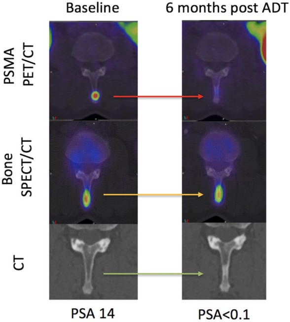
Figure 5.

Baseline and restaging 6 months following ADT in a patient with grade group IV prostate cancer. 68Ga-PSMA PET/CT (axial fused), SPECT/CT bone scan (axial fused) and CT scans centred on a spinous process osseous metastasis are shown. At baseline, the metastasis is seen on PSMA PET/CT and bone SPECT/CT but not CT. At 6 months, a complete biochemical response (PSA < 0.1 ng/ml) was achieved correlating with complete response on PSMA PET/CT. The bone SPECT/CT, however, was stable and the CT demonstrated a ‘new’ sclerotic lesion. The bone scan and CT are not true reflective of disease status at 6 months.
ADT, androgen-deprivation therapy; CT, computed tomography; PET, positron-emission tomography; PSA, prostate-specific antigen; PSMA, prostate-specific-membrane antigen; 68Ga-PSMA, gallium-68-labelled prostate-specific-membrane antigen; SPECT, single-photon-emission computed tomography.
