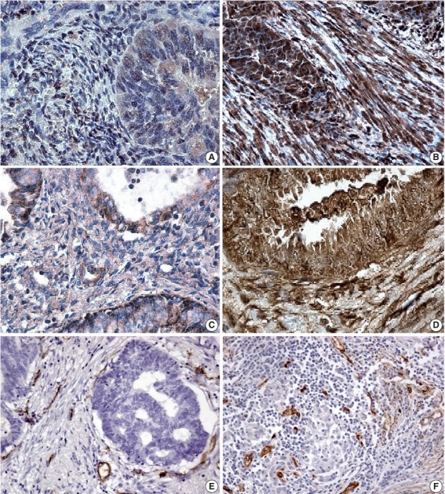
Fig. 2.

(A) A few positive glandular cells (microcystic, elongated and fragmented [MELF]–negative group). (B) All glandular cells positive for galectin-1 expression (MELF-positive group) in endometrioid carcinoma. (C) Positive local areas of cytoplasmic expression of vascular endothelial growth factor (VEGF; MELF-negative group). (D) Intense glandular and cytoplasmic (MELF-positive group) expression of VEGF in glands of the endometrioid carcinoma. Low (normal stroma) (E) and increased number of vessels (F) in fibromyxoid stroma situated around cancer cells forming gland without lumens.
