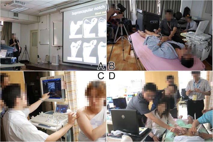
Fig. 2.
The course was divided into two parts. The didactic section consisted of (a) lectures based on six major joints and (b) standardization of scanning skills by using projections of ultrasound images and hand gestures on the screen in the auditorium room. The practical section comprised (c) demonstration of the sonoanatomy by the instructor and (d) scanning on the volunteer model by the attendee under the instructor’s assistance

