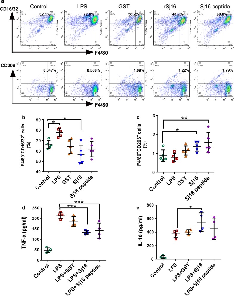
Fig. 5.
rSj16 protein and Sj16 peptide inhibited M1 macrophages and alternatively activated M2 macrophages in vitro. Peritoneal macrophages obtained from BABL/c mice were stimulated with LPS (1 μg/ml), GST (10 μg/ml), rSj16 (10 μg/ml), Sj16 peptide (10 µg/ml), or medium alone. Macrophages were harvested after 24 h. a The expression of CD16/32 (M1) and CD206 (M2) macrophages were evaluated by FCM analysis. b Percentages of F4/80+ CD16/32+ macrophages. c Percentages of F4/80+ CD206+ macrophages. d The expression of TNF-α. e The expression of IL-10. Results are expressed as the mean ± SEM of 3 independent experiments. Significant differences were detected, *P < 0.05

