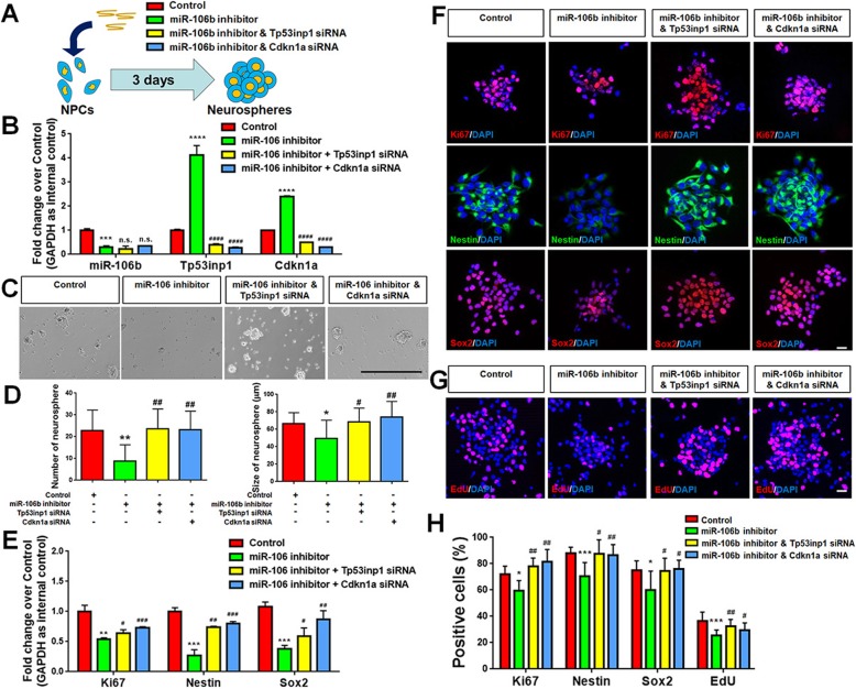
Fig. 6.
a–h miR-106b promotes the proliferation of NPCs through Tp53inp1-Tp53-Cdkn1a axis. a A schematic representation of the experimental approach. b qPCR analysis of expression levels of miR-106b and transcripts corresponding to Tp53inp1 and Cdkn1a in all experimental groups versus controls. c, d The number and size of neurospheres were quantified in all experimental groups versus controls. e qPCR analysis of levels of miR-106b and transcripts corresponding to Ki67, Nestin, Sox2, Tp53inp1, and Cdkn1a in all experimental groups versus controls. f Immunofluorescence analysis of transduced cells displaying proliferating cells (Ki67)- and NPCs (Nestin/Sox2)-specific immunoreactivities in all experimental groups versus controls. g Immunofluorescence analysis of transduced cells displaying EdU immunoreactivities in all experimental groups versus controls. h Quantification of cells expressing Ki67/Nestin/Sox2/EdU immunoreactivities in all experimental groups versus controls. Scale bar, 400 μm (b) and 20 μm (e, f). Data are mean ± SD. ∗∗∗∗p < 0.0001, ∗∗∗p < 0.001, ∗∗p < 0.01, and ∗p < 0.05. ####p < 0.0001, ###p < 0.001, ##p < 0.01, and #p < 0.05 versus the miR-106b LOF groups. Experiments were carried out three times in triplicates for in vitro perturbation

