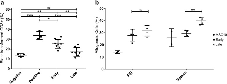
Fig. 7.
Immune inhibition of T-cells by early and late induced MSC-EMs. Early and late induced MSC-EMs were activated with IFN-γ (25 ng/mL) and TNF-α (1 ng/mL) for 4 days, washed with PBS, and used for conditioning of basic medium (MSC10) for 24 h. PBMC from HLA-mismatched donors were separately cultivated in the different media for 24 h. (a) For the mixed lymphocyte reaction assay, PBMC were mixed in a ratio of 1:1 and cultivated for 7 additional days. MSC10 was used as a negative control and the positive control contains IL2 (400 IU/mL). IL2 was also added to the conditioned media to activate T-cells. Conditioned medium of early and late induced MSC-EM inhibited blast transformation of T-cells. (b) For the in vivo killing assay, PBMC were stained with different colors, mixed medium wise, and injected in a 1:1 ratio in the respective media into mice previously reconstituted with a human immune system. Five days later, mice were sacrificed and analyzed by flow cytometry. Depicted are the percentages of allogeneic cells among all stained hCD3+ cells. Each point represents individual mice. Shown are the group means and standard deviation. Statistical tests were performed with ANOVA and p values calculated with Tukey’s multiple comparison. Only significant differences are indicated. ** = p < 0.01, *** = p < 0.001. PB = peripheral blood

