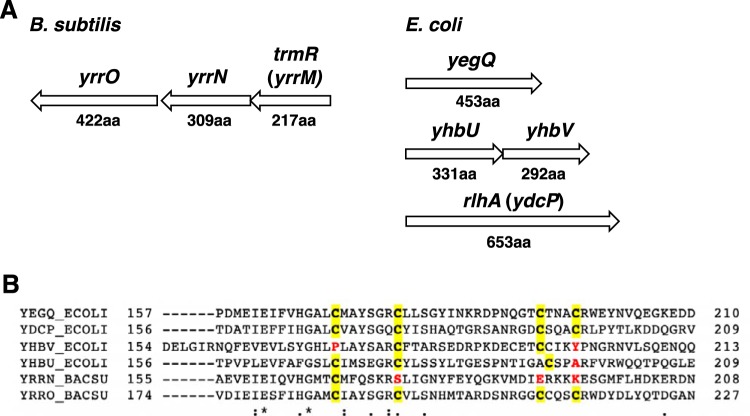
FIG 2.
(A) Organization of the two peptidase U32 genes in B. subtilis and four genes in E. coli. Numbers indicate corresponding protein amino acid length. (B) Clustal-omega amino acid alignment of peptidase U32 homologs from E. coli and B. subtilis. Putative Fe-S cluster cysteines are highlighted in yellow although there are other cysteines in the region. Amino acids that differ from cysteine in the conserved positions are shown in red (YhbU is the exception in which an adjacent cysteine has been included as a potential Fe-S ligand).

