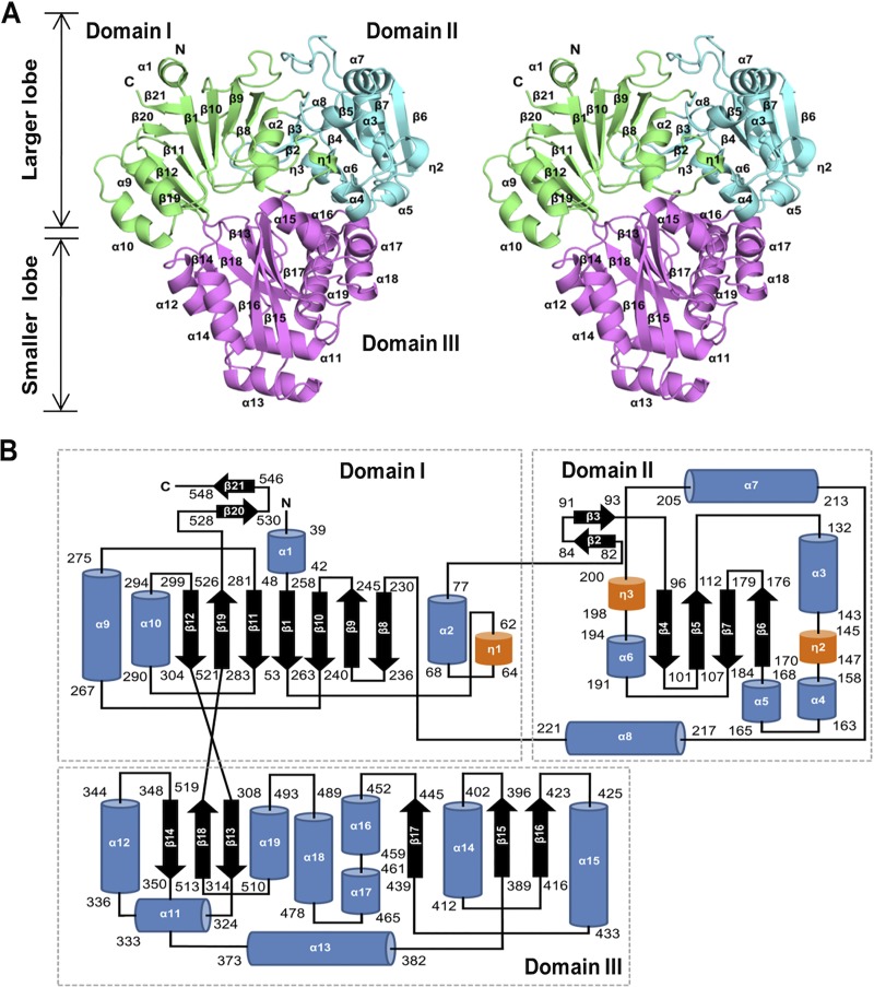
FIG 1.
Overall structure of H. pylori SS1 DppA. (A) Stereoview representation of the overall fold of HpDppA. The three domains (I, II, and III) are shown in green, cyan, and magenta, respectively, and labeled. (B) The topology of secondary structure elements of HpDppA. The α-helices are represented by blue rods, 310-helices are represented by orange rods, and β-strands are represented by arrows.

