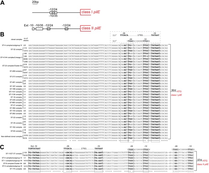
FIG 1.
Analysis of class I and class II pilE promoter regions in N. meningitidis. (A) Schematic representation of the class I and class II pilE promoter regions, based on N. meningitidis MC58 and 8013 (class I pilE) and FAM18 (class II pilE). An extended −10 and −10/−35 sequences are shown as open boxes, and the −12/−24 sequences are shown as striped boxes. (B and C) Clustal Omega alignment of the region upstream of pilE in isolates with class I (B) and class II (C) pilE. The clonal complex and the number of strains with the indicated sequences are shown. Sequences corresponding to −10/−35 and −12/−24 elements are boxed. E. coli consensus sequences (EσD −10/−35 and EσN −12/−24) are shown above the alignments. Asterisks indicate positions which have a single, fully conserved residue. Nucleotides in bold are identical to the consensus sequences.

