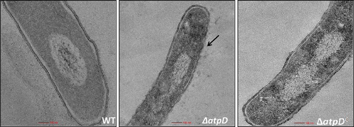
FIG 4.
Transmission electron microscopy-based ultrastructural analysis of M. smegmatis devoid of atpD. TEM micrographs of the WT, ΔatpD, and ΔatpDC strains (as indicated in each panel) are shown. The ultrastructure of the ΔatpD bacterium shows remarkable differences (marked with an arrow pointing to the altered cell envelope) from the other two. The experiments were performed at least thrice. Only one representative image is shown here. For more images, see Fig. S9 in the supplemental material.

