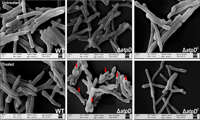
FIG 5.
Scanning electron microscopy-based assessment of the effect of SDS on the cell surface of the atpD-knockout strain. SEM images of the WT, ΔatpD, and ΔatpDC strains with or without treatment (as indicated in the images) with 0.1% SDS are shown. The pits formed in the case of the atpD-knockout due to SDS treatment are marked with arrows. The experiments were performed at least thrice; only one representative image is shown in each case. EHT, extra-high tension; WD, working distance.

