Figure 4.
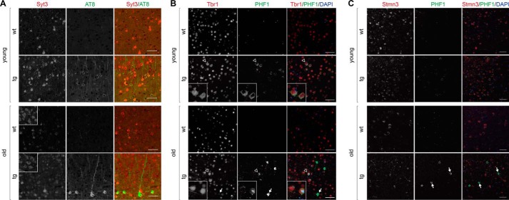
Altered expression of Syt3, Tbr1, and Stmn3 in tau harboring neurons of TAU58/2 mice. A, staining of young and old TAU58/2 and nontransgenic control brains with antibodies to Syt3 (red) and phosphorylated tau (AT8). Insets show Syt3 staining of dorsal thalamic nucleus as a reference area of same intensity in TAU58/2 and nontransgenic mice. Scale bar, 50 μm. B, staining of young and old TAU58/2 and nontransgenic control brains with antibodies to Tbr1 (red) and phosphorylated tau (PHF1). Merged images include nuclear DAPI (blue) staining. Arrows indicate NFTs with marked accumulation of phosphorylated tau and absence of Tbr1. Insets show higher resolution images of neurons indicated by open arrowheads in larger images. Scale bar, 50 μm. C, staining of young and old TAU58/2 and nontransgenic control brains with antibodies to Stmn3 (red) and phosphorylated tau (PHF1). Merged images include nuclear DAPI (blue) staining. Arrows indicate NFTs with marked accumulation of phosphorylated tau and absence of Stmn3. Scale bar, 50 μm.

