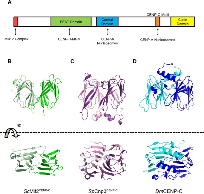
Figure 1.
Side and aerial views of CENP-C cupin domain crystal structures. A, classified domains of human CENP-C and their respective binding partners indicated by double-headed arrows. B, crystal structure of the ScMif2CENP-C cupin domain (PDB code 2VPV) (25). Monomers are colored green and light green. C, crystal structure of the SpCnp3CENP-C cupin domain (PDB code 6O2D). Monomers are colored in purple and pink. D, crystal structure of the DmCENP-C cupin domain (PDB code 6O2K). Monomers are colored in blue and cyan. Illustrations of protein structures used in all figures were generated with PyMOL (Delano Scientific, LLC). Both N and C termini are labeled.

