Figure 1.
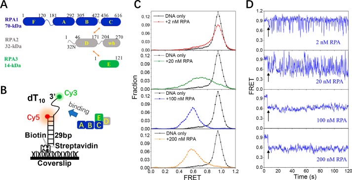
RPA undergoes dynamic association and dissociation with 10-nt ssDNA. A, molecular structure of RPA. wh in RPA2 represents the winged helix domain. Arrows indicate the intersubunit associations. B, schematic representation of the smFRET experimental setup. C, FRET histograms of the DNA substrate dT10 alone and with various concentrations of RPA. Each FRET histogram was constructed from more than 300 traces. D, representative FRET traces of dT10 in the presence of RPA. Black arrows indicate the addition of RPA.

