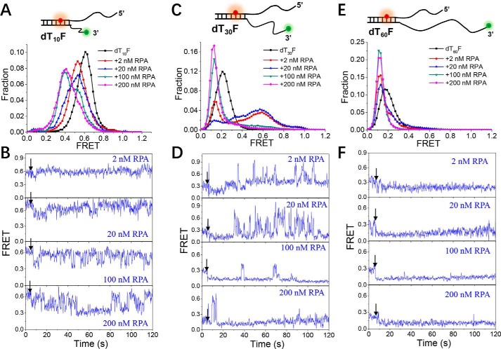
Figure 6.
The binding of RPA to the DNA fork structure is highly dynamic. A, C, and E, FRET histograms of the DNA substrates dT10F, dT30F, and dT60F alone and with various concentrations of RPA. Each FRET histogram was constructed from more than 300 traces. B, D, and F, representative FRET traces of dT10F, dT30F, and dT60F in the presence of 2–200 nm RPA. Black arrows indicate the addition of RPA.

