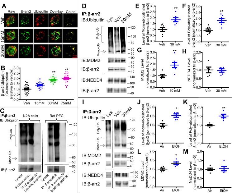
Figure 2.
Acute ethanol exposure increases ubiquitination of β-arrestin2 in N2A-5HT1AR cells and rat PFC. A, representative raw and binary fluorescent images of β-arrestin2 (green) and ubiquitin (red) and co-localization (Co-loc) in N2A-5HT1AR cells treated with ethanol (15–75 mm) or vehicle (Veh) (media) for 18 h. B, ethanol exposure dose-dependently increased β-arrestin2 co-localization with ubiquitin (n = 29–30 cells/group, six replicates, **, p < 0.01 versus vehicle, a one-way ANOVA followed by Bonferroni post hoc test). C, validation of the rabbit anti-β-arrestin2 antibody (LSBio; LS-B15546) was performed using a custom-made blocking peptide (GenScript; sequence DDIVFEDFARLRLK). Immunoprecipitation (IP) was performed using lysates from drug-naïve N2A-5HT1AR cells and rat PFC tissue in the presence and absence of the corresponding β-arrestin2 (β-arr)-blocking peptide (10 μg). Immunoprecipitates and N2A cell lysates were immunoblotted (IB) for mouse anti-ubiquitin (Santa Cruz Biotechnology; sc8017) and mouse anti-β-arrestin2 (LSBio; LS-B6008). β-Arrestin2–blocking peptide abolished immunoreactive bands detected in samples without the presence of this blocking peptide. D, representative blots for immunoprecipitated β-arrestin2, ubiquitin, and MDM2 from N2A-5HT1AR cell lysates. Cells were treated with ethanol (30 mm, 18 h) or vehicle (Veh) (media), and cell lysates were immunoprecipitated overnight with rabbit anti-β-arrestin2 antibody. Immunoprecipitates and cell lysates were immunoblotted with mouse anti-ubiquitin, mouse anti-MDM2, rabbit anti-NEDD4, and mouse anti-β-arrestin2 antibodies. E–H, quantification of β-arrestin2 mono- and poly-ubiquitination, MDM2, and NEDD4 (n = 5, **, p < 0.01 versus vehicle, unpaired Student's t test). I, representative blots for immunoprecipitated β-arrestin2, ubiquitin, and MDM2 in rat PFC lysates. Samples from ethanol (EtOH) and control (Air) exposed rats were immunoprecipitated with rabbit anti-β-arrestin2 overnight. Immunoprecipitates and tissue lysates were immunoblotted with the antibodies described above. J–M, quantification of immunoprecipitated β-arrestin2, ubiquitin, MDM2, and NEDD4 (n = 5, *, p < 0.05 versus vehicle, unpaired Student's t test).

