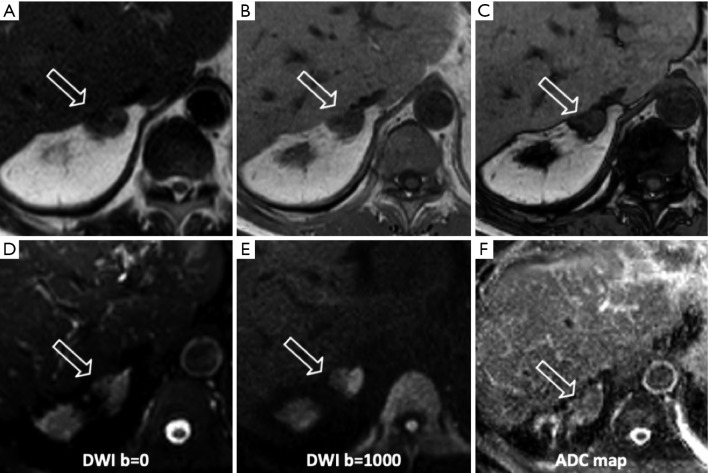
Figure 6.
A 70-year-old man with left adrenal metastasis from colon cancer. The lesion (arrows) shows signal hypointensity on the axial TSE T2-w (A) sequence without significant signal difference between the in-phase (B) and out-of-phase (C) sequences. Restricted diffusion is evident on b=1,000 DWI (D,E) sequences and ADC map (F). DWI, diffusion weighted imaging; ADC, apparent diffusion coefficient.

