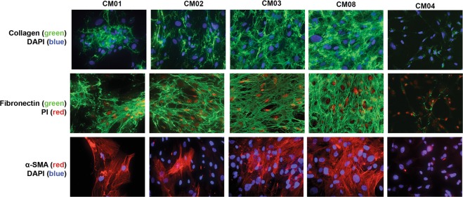
Fig. 6.
Immunocytochemical staining of cMASCs for ECM proteins collagen and fibronectin, and CAF marker α-SMA. DAPI or propidium iodide (PI) were used to visualize the nucleus. CM01, 02, 03, and 08-PDCs (cMASCs) showed abundant and intricate lattices of collagen and fibronectin and α-SMA staining was evident in a subpopulation of these cMASCs. By contrast, CM04-PDC had virtually no evidence of α-SMA, collagen or fibronectin expression

