Fig. 1.
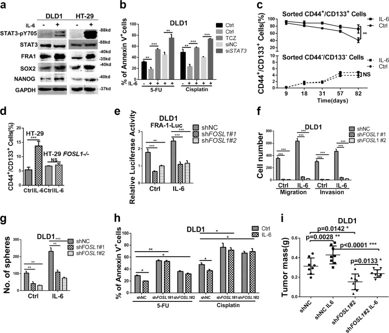
Interleukin (IL)-6 promotes colon cancer stemness in an FRA1-dependent manner. a Western blot analysis of DLD1 and HT-29 cells cultured in the presence/absence of 50 ng/ml IL-6 for 24 h. Protein levels of STAT3-pY705, STAT3, FRA1, SOX2, NANOG, and GAPDH were examined. b DLD1 cells were cultured in medium supplemented with the chemotherapeutic drugs 5-Fluorouracil (5-FU) and cisplatin and in the presence/absence of IL-6 (50 ng/ml), Tocilizumab (5 μg/ml), and siSTAT3. The percentage of apoptotic (Annexin V+) cells were employed as a measure of sensitivity to the treatment when compared to the controls. c CD44+/CD133+ and CD44−/CD133− DLD1 cells were sorted by fluorescence-activated cell sorter (FACS) and cultured in the presence/absence of IL-6 for the indicated times. These cultures were monitored at the indicated time points by FACS analysis. Quantification of CD44+/CD133+ cells in triplicate (mean ± SD) are depicted. d CD44/CD133 FACS analysis of parental (FOSL1+/+) and FOSL1−/− HT-29 cells cultured in the presence/absence of IL-6 for 7 days. The relative percentage of the CD44+/CD133+ subpopulation is indicated in the histogram. e FRA1 transcriptional activity was measured in shNC, shFOSL1#1, and shFOSL1#2 DLD1 cells cultured in the presence/absence of IL-6 (50 ng/ml). f, g Migration and invasion assays (d) and sphere formation assays (e) were performed with non-target control (shNC), shFOSL1#1, and shFOSL1#2 DLD1 cells cultured in the presence/absence of IL-6. Results are shown as histograms showing quantitative values of the number of cells (or spheres) from triplicate experiments (mean ± SD). h shNC and FOSL1 knockdown (shFOSL1#1 and shFOSL1#2) DLD1 cells were cultured in medium supplemented with the chemotherapeutic drugs 5-FU and Cisplatin and in the presence/absence of IL-6 (50 ng/ml). The percentage of apoptotic (Annexin V+) cells indicated that both shFOSL1#1 and shFOSL1#2 were characterized by increased sensitivity when compared to the shNC control cells. i 5 × 10⁵ shFOSL1#2 and, as a control, shNC DLD1 cells were cultured in the presence/absence of IL-6 for 5 days and then injected subcutaneously into the flanks of BALB/C nude mice. DLD1 cells treated with IL-6 increased tumor mass when compared with the shNC group. And this effect was attenuated upon FOSL1 knockdown. *p < 0.05, **p < 0.01, ***p < 0.001. Unpaired t test. Data are presented as mean ± SD

