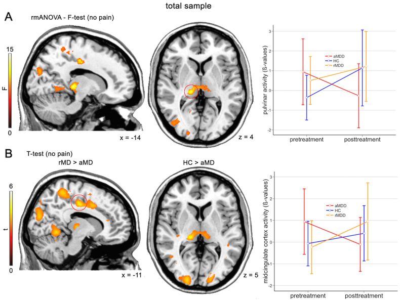
Fig. 2.
Reversal of thalamic hyperactivity after antidepressant treatment. Repeated measures analysis of variance (rmANOVA) comparisons between fMRI-1 and fMRI-2 within healthy controls (HC), remitted depressed patients (rMD), and acute depressed patients (aMD). Acute patients were treated with escitalopram or venlafaxine for 12 weeks. a, F-test results depicts elevated activation of thalamic pulvinar nuclei in acute patients, which reversed to baseline values of HC after treatment. Activations represents difference in F-test between fMRI-2 vs. fMRI-1. b, Post-hoc t-tests revealed elevated activity in midcingulate cortex. Activations represents difference in t-test between fMRI-2 vs. fMRI-1. Errorbar-plots represent activation values of red circled-clusters, which survived family-wise error correction (FWE, cluster-level, p < 0.05). Left is left. Results are depicted at p < 0.001 uncorrected voxel level

