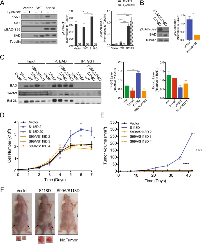
Fig. 3.
BAD S99 phosphorylation is required for S118D-mediated cell and tumor growth. a Left: MDA-MB-231 cells expressing pcDNA3.2-V5-DEST vector control, WT-BAD, or BAD-S118D were grown normally for 3 days prior to 50 μM Ly294002 addition for 1 h prior to cellular lysis. Right: Quantification of protein band density (error bars ± SEM of 3 independent experiments). b Left: Immunoblot depicting BAD protein levels in indicated cell lines. Right: Quantification of protein band density (error bars ± SEM of 3 independent experiments). c–f MDA-MB-231 cells expressing pcDNA3.2-V5-DEST vector control, BAD-S118D, BAD-S118A, and BAD S99A/S118D. c MDA-MB-231 cells expressing the indicated mutations were subjected to immunoprecipitation by BAD or GST (control) antibodies and immunoblotted against BAD, 14-3-3, and Bcl-XL. Levels of 14-3-3 and Bcl-XL binding were compared to BAD IP levels (error bars ± SEM of 3 independent experiments). d Cell count assay over 7 days (error bars ± SEM of 3 independent exkperiments). e A total of 3 × 10⁶ cells of vector, BAD-S118D, or BAD-S99A/S118D, were injected into the subcutaneous flanks of nude mice. Tumor volume was measured weekly for 7 weeks (vector n = 4, BAD-S118D n = 6, BAD-S99A/S118D clone 2 n = 6, clone 3 n = 4, clone 4 n = 5; error bars ± SEM). f Representative images of subcutaneous tumor growth of nude mice taken at day 42. Black arrows indicate tumor location. Images of tumors are depicted below. Scale bar = 0.5 cm

