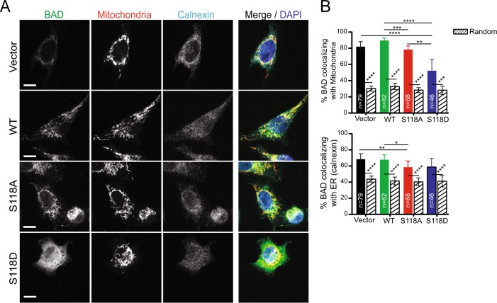
Fig. 4.
Wild-type BAD co-localizes strongly with mitochondria. a Z slice of confocal imaging of MDA-MB-231 cells expressing pcDNA3.2-V5-DEST vector control, WT-BAD, BAD-S118D or BAD-S118A immunostained with antibodies against BAD (green) and calnexin (endoplasmic reticulum (ER) marker) (cyan). MitoTracker™ Red and DAPI were used as mitochondria (red) and nuclei (blue) markers, respectively. Scale bar = 10 μm. b Percentage of BAD protein (BAD channel MCC fraction converted to percentage) co-localizing with mitochondria (top) and ER (bottom) (colored bars) compared to randomized control (patterned bars) (n = number of cells; Kruskal-Wallis test)

