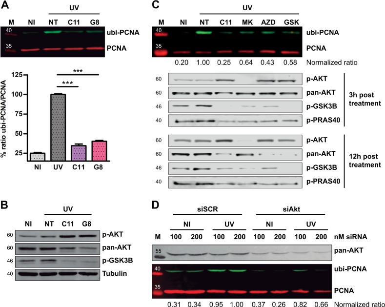
Fig. 2.
AKT inhibition impairs PCNA ubiquitylation. a U2OS cells were UV irradiated (15 J/m2) and treated for 12 h with the indicated inhibitors at 1 μM. The western blot shows the strong PCNA ubiquitylation inhibitory activity found in two structurally related hits: C11 (compound #: GSK1581428A) and G8 (compound #: GSK1389063A). The graph in the lower panel shows the quantification of three independent experiments. Statistical analysis was performed using analysis of variance (ANOVA) with Tukey Kramer post-test (***p ≤ 0.001). b U2OS cells were treated as in a and western blots with specific antibodies were performed to study pAKT, total AKT, and p-GSK3β levels. α-Tubulin was used as a loading control. c U2OS cells were pre-treated for 12 h using 0.5 µM C11 and 5 µM of the structurally unrelated AKT inhibitors: MK-2206 (Merck), AZD5363 (AstraZeneca), GSK690693 (GlaxoSmithKline). After UV, all these inhibitors were used at 20 µM and C11 was used at 1 µM. The normalized ubi-PCNA/total PCNA ratios are shown below the PCNA panel. pAKT, AKT, p-GSK3β, and p-PRAS40 western blots were performed at 3 and 12 h post-treatment to confirm the AKT inhibitory activity of each compound. d U2OS cell were transfected with two concentrations of siRNAs. Forty-eight hours later, cells were UV irradiated, and after 12 h, samples were processed for quantification of PCNA ubiquitylation by western blot. A western blot for pan-AKT was performed to confirm the siRNA-mediated knockdown. The normalized ubi-PCNA/total PCNA ratios are shown below the PCNA panel

