Fig. 7.
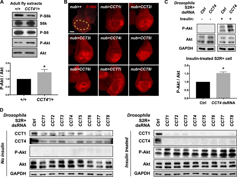
Reduced CCT complex affects the level of phosphorylated S6K and Akt. a Western blot of wild type and CCT4KG09280/+ adult extracts. Phospho-S6K and phospho-S6 levels were reduced in CCT4KG09280/+. Total S6K and Akt levels were not changed. P-Akt levels normalized to total Akt levels were shown in a bar graph. *P < 0.05. n = 3. b Increased phospho-Akt in CCT-knockdown wing discs. RNAi knockdown of each CCT gene using nub-Gal4 increased P-Akt level compared to nub>+. Nub-Gal4-expressing area is marked by yellow dashed line. Scale bar, 100 μm. c CCT4 RNAi increases the P-Akt level in insulin-treated S2R+ cells. P-Akt levels were increased in insulin-treated S2R+ cells. Insulin (25 µg/ml) was treated for 10 min. GAPDH levels were used as loading controls. P-Akt levels normalized to total Akt levels in insulin-treated S2R+ cells were shown as graph. *P < 0.05. n = 3. d The level of phospho-Akt in CCT-knockdown S2R+ cells. When insulin (25 µg/ml) was treated for 10 min, P-Akt levels were increased in each CCT RNAi cell lysate. GAPDH levels were used as loading controls

