Fig. 3.
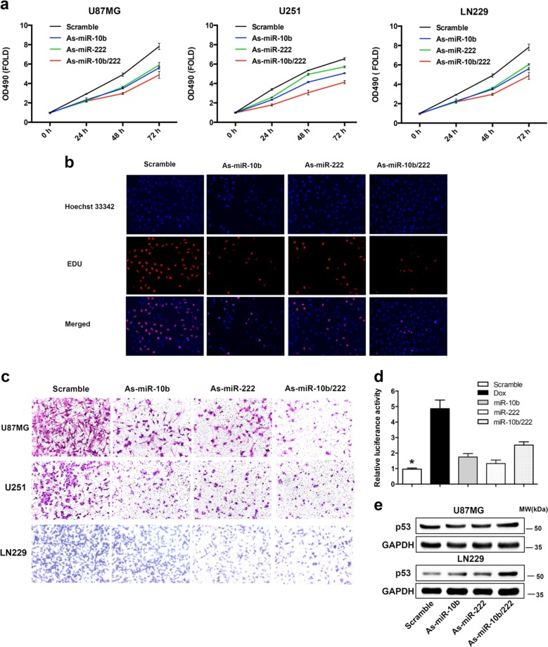
Inhibition of miR-10b and miR-222 suppressed the GBM cells proliferation and invasiveness by activation of p53. a LN229, U87MG, and U251 cells were transfected As-miR-10b or/and As-miR-222 and harvested after 24, 48, and 72 h. Cell viability was analyzed by CCK‐8 assay. b Representative results of the EdU proliferation assay of LN229. All cells were transfected with miRNA inhibitors and labeled with EdU (red), Hoechst 33342 (blue). c Representative images of Transwell assays of cells after transfection with miRNA inhibitor. d LN229 cells were co-transfected with pGL4.38 [luc2P/p53 RE/Hygro] and pGL4.74 [hRluc/TK] vector plasmids. 24 h after transfection, the cells were treated with miR-Scr, As-miR-10b, As-miR-222, and As-miR-10b/222 for 24 h. Cells were only stimulation with doxorubicin (5 mM) for 18 h as positive control. The data were represented as mean ± s.d. (n = 3), *p < 0.01 versus scramble group. e Western blot analysis showed the level of p53 after inhibition of miR-10b and miR-222 in LN229 and U87MG. GAPDH was used as an internal control. The RT-PCR results for TP53 mRNA in LN229 and U87MG were show in Supplementary Fig. S2

