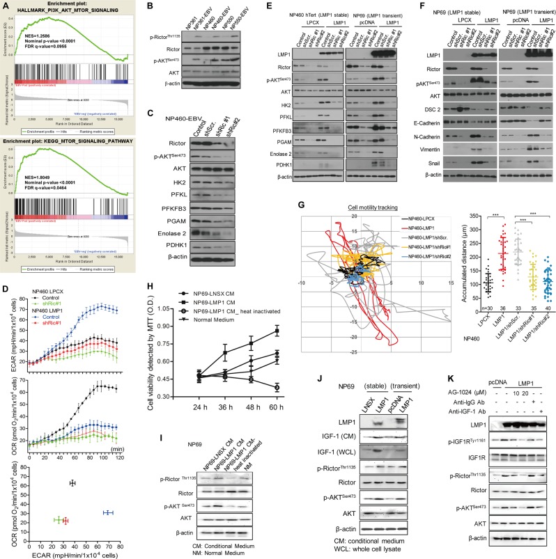
Fig. 3.
LMP1 activation of mTORC2 by autocrine secretion of IGF1 links Epstein-Barr virus (EBV)-reprogrammed glucose metabolism to cell motility. a GSEA showing the EBV infection upregulate PI3K-AKT-mTOR signaling-dependent gene sets. In each panel, the top portions of the plots show the ES for the gene set. Each vertical bar in the middle portions represents a gene, and genes enriched in either condition are at the right (EBV-positive) or left (EBV-negative) parts of the graph. The NES, the p value, and the false discovery rate (q-value) are indicated in the insert. b Different NPE cell lines with or without EBV infection, and c NP460-EBV cell infected with shRictor or control empty lentiviral vector were subjected to western blotting for detecting the expression of mTORC2 activity and metabolism-associated molecules using specific antibodies. β-actin expression was used as the loading control. d NP460-LPCX and NP460-LMP1 cells infected with shRictor or control empty lentiviral vector were plated at 10,000 cells/well for 24 h, then the cells were incubated with the ECAR or OCR reagents according to the manual. ECAR and OCR were measured simultaneously by using the 96-well plate reading system (Victor, PerkinElmer) in real time. e, f NPE cells with stable or transient overexpression of LMP1 were infected with shRictor or control vector. Then, cells were lysed and analyzed by western blotting. g shRictor- or its control vector-infected NP460-LPCX and LMP1 cells were seeded on the coverglass chamber and observed under time-lapse microscope. Representative tracks of cell movements were traced and visualized using metaphase software every 10 min for 24 h. The accumulated distance was analyzed by metaphase software. h Cell viability of NP69 was measured by MTT assay after incubating with different conditional medium for the indicated times. i The NP69 cells were incubated with the different conditional medium for 48 h, followed by western blotting for detection of mTORC2 signaling activity using the indicated antibodies. j After culturing different pairs of NPE and NPC cells for 48 h, the culture media as well as the cell lysates were subjected to western blot assay using the indicated antibodies. k NP69-pLNSX and LMP1 cells were treated with different doses of AG-1024 and antibodies against-IgG or IGF1 for indicated time, followed by western blot assay using the indicated antibodies. Data are means ± SD (means ± SEM for g). ***p < 0.005

