Fig. 3.
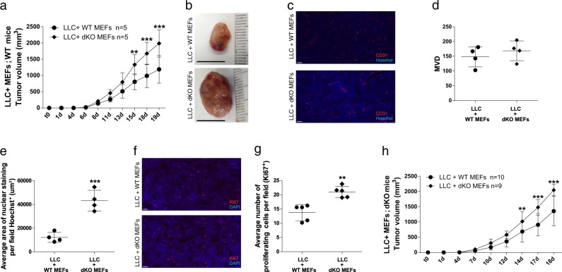
dKO MEFs promote LLC tumor growth, cell proliferation and blood vessel perfusion. a WT mice were subcutaneously implanted into the flanks with Matrigel containing cell mixture composed of LLC cells (0.5 × 10⁶ cells per mouse) together with MEFs (1.5 × 10⁶ cells per mouse) derived from either WT or dKO mice. Data are presented as mean ± SD, Two-way repeated-measures ANOVA followed by Bonferroni post-tests, n = 5, **p < 0.01; ***p < 0.001. Representative image of at least three experiments. b Representative images of tumors from a. c Representative images of tumor sections from a, stained with anti-CD31 (endothelial cells; red) and with Hoechst (nuclei; blue). Scale bar, 100 µm. d, e Quantifications of microvessel density (MVD, d) and nuclear staining (e) per field. Each dot represents the mean of 5 fields taken from one mouse. Data are presented as mean ± SD, Student’s t-test, n = 4, ***p < 0.001. f Representative images of tumor sections from a, stained with anti-Ki67 and DAPI. Scale bar, 50 µm. g The number of proliferating cells (Ki67+) per field. Each dot represents the mean of 5 fields taken from one mouse. Data are presented as mean ± SD, Student’s t-test, n = 5, **p < 0.01. Representative images of at least three experiments. h dKO mice were subcutaneously implanted as described in a, and tumor volume was monitored over time. Data are presented as mean ± SD, Two-way repeated-measures ANOVA with Bonferroni post-tests, n = 10,9, **p < 0.05; ***p < 0.001. Representative image of two experiments

