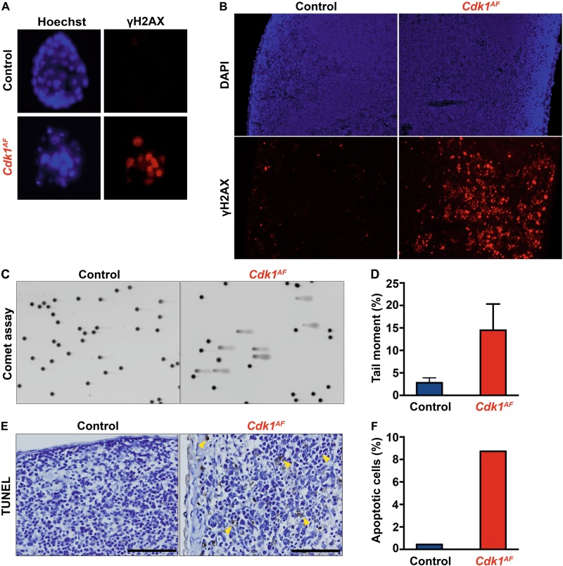
Fig. 1.
The expression of CDK1AF leads to early embryonic lethality. a Control (Cdk1+/SAF) and β-actin-Cre Cdk1+/AF blastocysts were visualized with Hoechst staining (nuclei). The level of DNA damage was assessed through immunofluorescence staining of phospho-H2AX. b The expression of Cdk1+/AF was induced in all tissues of adult Rosa26-CreERT2 mice upon tamoxifen IP administration. Spleen sections were stained for phospho-γH2AX to visualize DNA damage response. c DNA damage in spleen of control and Rosa26-CreERT2 mutant mice was analyzed by Comet assays using the tail moment as a parameter to determine the extent of DNA breaks. d Quantification of DNA breaks was calculated based on the tail moment. e To investigate chromosomal fragmentation, spleen sections from tamoxifen injected control and Cdk1+/AF Rosa26-CreERT2 mice were analyzed by TUNEL assay to evaluate apoptosis. Yellow arrows indicate apoptotic cells with extensive DNA breaks. f The number of apoptotic cells was quantified in relation to the total number of cells

